THIS ARTICLE/PRESS RELEASE IS PAID FOR AND PRESENTED BY University of Oslo - read more
A new treatment can save the eyesight of those who get a serious eye infection
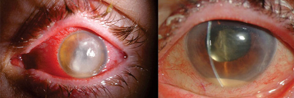
Some patients develop a serious infection in the eye after eye surgery. The risk of becoming blind is high without prompt treatment. A new approach to diagnostics and treatment can help save patients’ vision.
Fortunately, eye surgery for conditions such as cataract is successful in most cases. Even so, in some cases, bacteria can enter the eye during surgery and cause a serious infection in the eye called endophthalmitis.
Endophthalmitis is the opthalmologists' most feared complication. In the worst case, the infection can cause blindness.
It is therefore crucial to diagnose and treat the infection as soon as possible. The treatment consists of antibiotics as well as a surgical intervention to remove pus from the eye.
Ophthalmologists also take samples from the inside of the eye for microbiological analysis. Usually, patients have samples from the eye taken before they receive antibiotics.
However, a new study shows that it is possible to do this in the opposite order. In other words, to give antibiotics immediately when ophthalmologists suspect that the patient has an infection in the eye.
Receiving antibiotics more rapidly can help save patients’ vision.
"We found that 7 out of 10 endophthalmitis patients achieve a significant improvement in vision when they receive antibiotic treatment straight away, and the samples are taken afterwards," PhD candidate and ophthalmologist Kathrine Blom says.
She is in the process of completing her doctorate at the Faculty of Medicine at the University of Oslo (UiO) and is employed at the Department of Ophthalmology at Oslo University Hospital.

PCR technology makes immediate treatment with antibiotics possible
Most people heard about PCR tests during the Covid-19 pandemic. A variant of the same technology is also the key to success in the endophthalmitis study.
Analysing the samples from the eye with PCR technology made it possible to give patients antibiotics immediately.
PCR is a technology that identifies the genetic material of bacteria and viruses, regardless of whether these are alive or dead. It therefore does not matter whether the samples are taken before or after the patient has received antibiotics, which have possibly killed the bacteria.
"Securing pus samples from the inside of the eye is not easy, as it requires both surgical expertise and advanced equipment. In fact, it is much easier to inject antibiotics. Prioritising immediate antibiotic treatment over diagnostics thus means that we can treat patients with endophthalmitis quicker than before. Immediate treatment improves the prognosis," Blom explains.
Need to know which bacteria are causing the eye infection
The samples and microbiological analysis are necessary in order to diagnose and treat the eye infection in the best possible way.
"We treat the eye infection with a combination of antibiotics that we know from experience work well against all common bacteria. However, the composition of bacteria can change over time," the researcher says. "It is therefore important to have a good overview of the variants of the bacteria that cause endophthalmitis. We can then adjust the treatment and give other types of antibiotics if necessary."
She points out that, historically, cataract surgery has been the most frequent cause of the infection.
"However, this is changing. Today, the most common cause is injection treatment of eye diseases with biological medicines, so-called anti-VEGF treatment. We see a slightly different composition of bacteria in these eye infections than those following cataract surgery," Blom says. "The more we know about the bacteria that cause the infection, the better we can adapt the treatment."
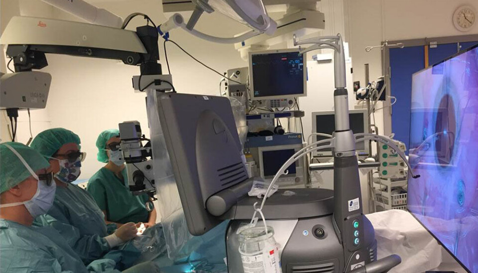
Make three small holes through the eye wall
The PCR samples are taken in connection with the surgical removal of the vitreous body of the eye, which fills the eyeball. The procedure is called a vitrectomy. The patient receives local anaesthetic.
Ophthalmologists then make three small holes through the eye wall.
"Using special instruments, we can then remove the pus and take samples at the same time. So this procedure involves both diagnostics and treatment," Blom explains.
The empty space in the eyeball is then filled with saline, air or gas, in addition to antibiotics.
Bacterial culture in the laboratory
The most common way to look for the bacteria has been to use a method called 'microbiological culture'. Culturing involves letting the bacteria reproduce in dishes in a laboratory.
However, to perform a bacterial culture, the bacteria must be alive. Since antibiotics kill sensitive bacteria, the samples must be collected before antibiotic treatment for this method of analysis to be used.
PCR technology, where it does not matter whether bacteria are dead or alive, thus offers new opportunities.
"PCR is a more sensitive method than culture. We detected more disease-causing endophthalmitis bacteria with PCR than with culture," the researcher says.
This was the case regardless of whether the samples were taken before or after the patient received antibiotics. At the same time, the traditional cultivation method also has an advantage.
"Culture can demonstrate whether the bacteria are sensitive or resistant to antibiotics, i.e., whether the antibiotics will be able to kill the bacteria or not. PCR cannot yet tell us this. We therefore think that it is best to combine both methods," Blom explains.
"The study is a milestone in the treatment of endophthalmitis"
Professor Ragnheidur Bragadottir at the Department of Ophthalmology at the Institute of Clinical Medicine and Oslo University Hospital is one of Kathrine Blom's supervisors. She refers to the study as a milestone.
"The study is a milestone in the treatment of endophthalmitis. It is the first prospective study of its kind internationally," the Professor says.
Close collaboration has been important to be able to carry out the study.
"I would like to thank the Department of Microbiology at Oslo University Hospital Ullevål for our collaboration. They have made it possible for us to take PCR in addition to culture on all endophthalmitis patients. We also wish to thank the Norwegian Association of the Blind and Partially Sighted – NABP for their financial contribution," Blom says.
Reference:
Blom et al. Primary vitrectomy or intravitreal antibiotics followed by early vitrectomy for acute endophthalmitis: A prospective observational study, Acta Ophthalmologica, 2022. DOI: 10.1111/aos.15207

This article/press release is paid for and presented by the University of Oslo
This content is created by the University of Oslo's communication staff, who use this platform to communicate science and share results from research with the public. The University of Oslo is one of more than 80 owners of ScienceNorway.no. Read more here.
See more content from the University of Oslo:
-
The likelihood of relationship breakdown may be influenced by our genes
-
Technology is making the music industry more individualistic and less equal
-
Researchers: Digital ID management in Norway is a catastrophe
-
Archaeology is helping researchers understand the effects of forced displacement
-
For 'Amalie,' going to prison was devastating. But it helped her get sober
-
“My pulse quickens every time someone says that children and young people are digital natives"


