THIS ARTICLE/PRESS RELEASE IS PAID FOR AND PRESENTED BY NTNU Norwegian University of Science and Technology - read more

The tiny structures that mimic a cell’s natural environment
Covering glass microscope slides in tiny, nano-sized pillars can mimic a cell’s natural environment – and could help biologists understand how cells act inside the human body.
When biologists study cells under a microscope, they look at them on flat surfaces that are nothing like the environment inside the human body. Now, researchers at NTNU have found a way to mimic some aspects of a cell’s native environment using tiny polymer pillars. Their work, funded by the Research Council of Norway, is published in the journal Nanoscale Research Letters.
“Cells in the human body are embedded in a complex matrix of molecules,” says Pawel Sikorski, a professor in the Department of Physics at NTNU. This environment – known as the extracellular matrix – is a dynamic support network for cells, providing not just physical scaffolding for tissues and organs to grow but also conveying signals to help cells communicate with each other. While taking cells out of the extracellular matrix and putting them on flat surfaces made of glass allows researchers to study them in the lab, it does mean we could be missing out on observing many cellular processes.
“Glass is very hard and the cell will sense that the substrate does not deform as it tries to pull on it,” says Sikorski. “That induces certain types of behaviour and also induces certain types of processes in the cells. They will behave differently if they were placed on something which is elastic and soft and can be deformed and remodelled.”
This means that if researchers want to understand how cells behave in their native environment, they need a substrate which replicates biology more closely. Embedding cells within hydrogels – for example, 3D networks of gelatin-like polymers – is one option. But studying cells within a hydrogel isn’t as easy as looking at them on a simple glass slide under an optical microscope. “If you want to see what’s happening it gets quite challenging,” says Sikorski.
Creating structures in a thin polymer film
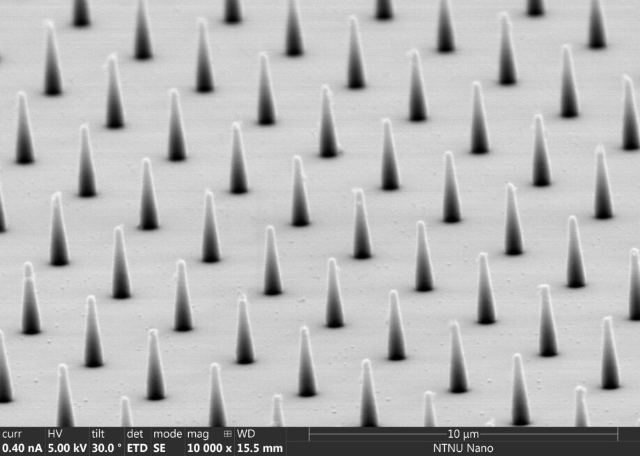
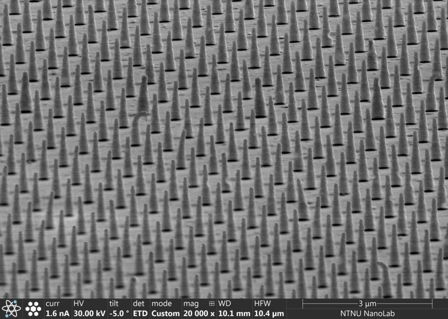
Mimicking some of the mechanical aspects of softer substrates with nanostructures is one possible way to address this problem – and that’s exactly what Sikorski and PhD student Jakob Vinje have done, in collaboration with cell biologists Noemi Antonella Guadagno and Cinzia Progida at the University of Oslo. Vinje covered glass slides in tiny pillars made of a polymer known as SU-8. These nanopillars – each measuring just 100 nanometres across at the tip – were made using electron beam lithography at NTNU NanoLab, where a focused beam of electrons creates structures in a thin polymer film.
“Per millimetre square you already have quite a lot of pillars, and if you want to study cells, then we need to make surfaces which are at least on the order of 10 by 10 millimetres,” says Sikorski. “The tools in NTNU NanoLab are essential for this to be possible.”
The researchers created substrates with a variety of different nanopillar arrangements and tested them using cells which produce fluorescent proteins. Looking at the cells under a microscope, the researchers analysed the shape, size and distribution of the points at which the cell attaches to the different surfaces.
Tightly packed pillars
After making hundreds of observations of cells on the various surfaces, the researchers found that substrates with tightly packed nanopillars most closely mimicked a softer surface. “If we make a substrate with dense pillars then the cells behave as if they were on a much softer substrate,” says Sikorski.
The beauty of the nanopillar-covered substrates is their simplicity – in theory, biologists could simply swap their usual glass slides for the new ones. “It has more features and more tuneability than a glass substrate, but it’s still relatively simple,” says Sikorski.
He says the ultimate aim would be for researchers to be able to “just open the package and take one of them out, put their cells on, study it under the microscope and then throw it away once they’re done.” However, for that to become a reality the substrates would need to be produced in their hundreds at a relatively low cost.
So far the researchers have only made a small number of prototypes, but there are existing methods – such as a low-cost, high-throughput technique for making nanoscale patterns called nanoimprint lithography – that could make scaling up production of the substrates possible.
As well as allowing biologists to study cells in a new way, the substrates could be used to develop better ways to screen medicines. To find a drug that stops cells sticking to a particular surface, for example, a nanopillar-covered substrate could mimic that surface and put potential medicines to the test.
Reference:
Jakob B. Vinje et.al.: Analysis of Actin and Focal Adhesion Organisation in U2OS Cells on Polymer Nanostructures. Nanoscale Res Lett, 2021.

See more content from NTNU:
-
New findings about the evolution of the brain: "I was blown away. My first reaction was, is this real?
-
Researchers are surprised that these chemicals have skyrocketed in Svalbard
-
Why some people can’t stop pulling out their hair or picking their skin
-
Can Europe create AI that we actually understand?
-
Doctors and nurses believe their own substance use affects patients
-
When the offshore wind turbines fall silent: What happens to the waste?


