An article from University of Oslo
Cutting the liver piece by piece
New surgical methods give hope to patients with cancer that has spread from the intestine to the liver. The disease can be changed from terminal to chronic by cutting the liver piece by piece using keyhole surgery.
Denne artikkelen er over ti år gammel og kan inneholde utdatert informasjon.
Each year, 3500 Norwegian develop colorectal cancer. Most of them undergo surgery. In half of them, the cancer spreads. Thirty per cent of those in whom the cancer has spread can now be operated on with a new surgical method.
“With our new surgical method we can transform an acute, terminal disease into a chronic disease,” says one of the world’s leading surgeons in the field of laparoscopy (keyhole surgery), Professor Bjørn Edwin at the Intervention Centre of UiO and Oslo University Hospital.
His solution is to preserve as much of the liver as possible.
“We try to take as little as possible. This is a completely new way of thinking.”
The most common surgical method is still to remove half of the liver if the cancer has spread to parts of it.
“By removing only a small piece at a time, we increase the chances that the patient can have repeat surgery. In other words, we try to preserve the liver to have some of it left if the cancer should return. Most of the patients have a recurrence within ten years. The idea is this: If we leave parts of the right side, we can later remove the left side of the liver. This is what we refer to as good liver housekeeping,” Bjørn Edwin explains.
He notes that the method only works for patients in whom the cancer has spread from the intestine to the liver. For patients with a primary tumour in the liver, other treatment methods are used.
For the last twenty years, Edwin has remained one of the country’s leading pioneers in the field of laparoscopy.
“With laparoscopy, we can intervene and operate many times, since the method does not produce the same adhesions as open surgery does. The results are equally good, and there are fewer complications. In addition, laparoscopy has taught the surgeons to operate more neatly,” Bjørn Edwin states.
Four small holes
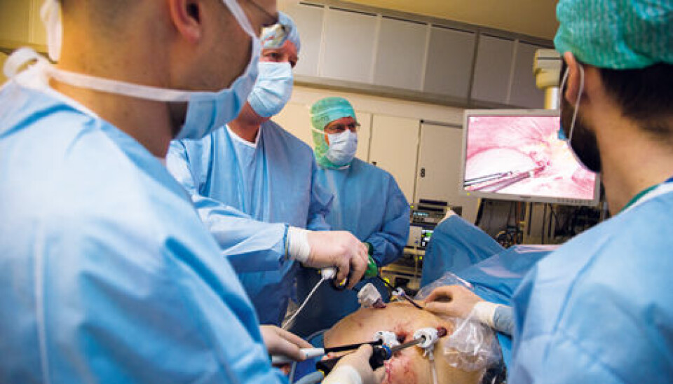
This way to perform surgery is far better for the patient than a classic operation. Instead of cutting open the abdomen, the surgeon makes four small holes. He can then use trocars, which are stiff tubes, to insert his instruments and a camera. One hole is for the video camera.
Through the other holes, the surgeon can introduce a forceps, an instrument, a scalpel or other implements needed to cut the liver loose. Before starting the procedure, however, they must first inflate the abdomen to provide a large cavity where they can work.
A large amount of blood flows through the liver. To avoid haemorrhages, the blood flow to the part which is to be operated on must be halted by ligating the arteries. This can be done by tying a suture around the blood vessels.
“If the patient nevertheless should start haemorrhaging we must switch to a regular operation. This happens in only one of a hundred cases. Previously, we also opened the abdomen occasionally to be on the safe side.”
Many different techniques can be used to cut the liver. The surgeons can use a scalpel, ultrasound or electrosurgical forceps. To incise the large blood vessels they use a sewing machine. Before the blood vessel is cut, they block it by sewing three rows of clips on each side of the cut.
The excised section of liver must be withdrawn from the body intact.
If the excised section of liver were ground up, it could be brought out of the body in liquid form, but then the pathologists would be unable to analyse the tumours. The liver section must therefore be removed whole.
This is done by inserting a plastic bag through one of the stiff tubes. The bag has the shape of an angler’s hand net.
“We catch the liver, close the bag, pull it towards the skin and out through a small incision.”
A gentle procedure
Laparoscopy is not only less painful than classic surgery, but the patients recover more quickly and spend fewer days in hospital.
“The average hospitalization period after a laparoscopic intervention is three days, while patients spend five to seven days in hospital after open surgery.”
Edwin is currently investigating the correlation between surgical methods and survival rates.
“Based on the illness histories of patients who have been operated on for cancer that has spread to the liver, expected survival is higher with laparoscopy, but the responses are not statistically significant. We don’t know exactly why the survival rates increase, but it might be related to the immune response.”
The story behind
Gastric surgeons started to use keyhole surgery in the late eighties, initially on gall-bladder patients. This surgical method was described one hundred years ago, and was first used by gynaecologists during the forties.
“In those days, the surgeon looked through a telescope, but having video on a screen enabled us to perform laparoscopy in a far more sophisticated way.”
Bjørn Edwin is a pioneer in the field of keyhole surgery and was among the very first to use this method for liver surgery. Now, Bjørn Edwin has taught his method to surgeons in Norway as well as abroad.
“Laparoscopy will increasingly replace open surgery. Ten years after we started, other countries have followed. American hospitals are still opposed to the surgical method for preserving liver tissue. France is now using it, and Russia is following suit.”
When he attended a world conference on cancer of the liver and pancreas in 2008, hardly anyone used laparoscopy on these organs. Two years later, there were some. At this year’s conference in Seoul it became evident that many have now started to use this new surgical technique.
Interdisciplinary team
To perform modern laparoscopy, collaboration with the engineers is absolutely essential. Using a three-dimensional map of the liver, the surgical team can see where the blood vessels are located and plan where to make their incisions accordingly.
“We are now undertaking research to enable us to see the movements of the instruments interactively on the map of the liver during surgery,” Bjørn Edwin says. He emphasizes that the trials with modern laparoscopy would have been impossible without the strong interdisciplinary team at the Intervention Centre.
“There, we have close proximity to physicists, radiologists and other specialists. It’s a superb setting for undertaking a development stage,” Edwin points out.