An article from University of Oslo
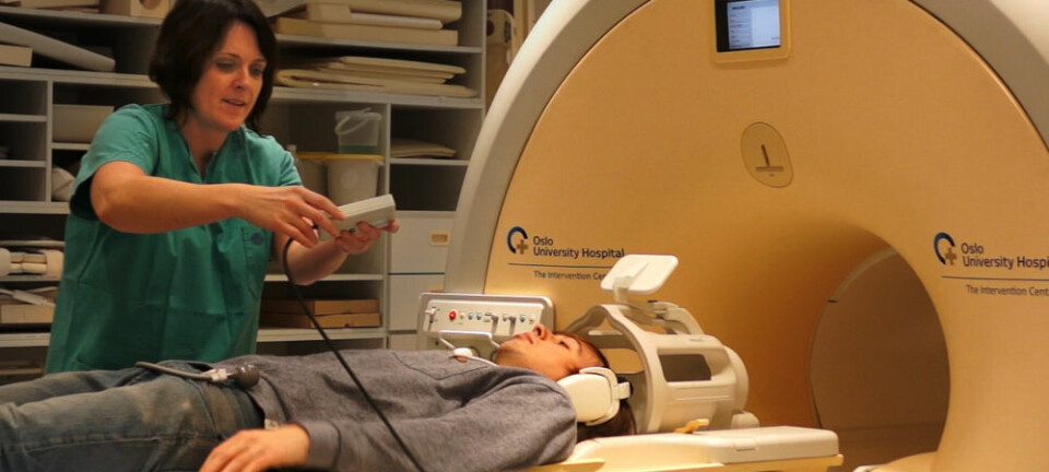
A pipette in brain tissue
The activity within a synapse of the brain has been measured for the very first time in Scandinavia, opening up new doors to our knowledge about learning and memory.
Denne artikkelen er over ti år gammel og kan inneholde utdatert informasjon.
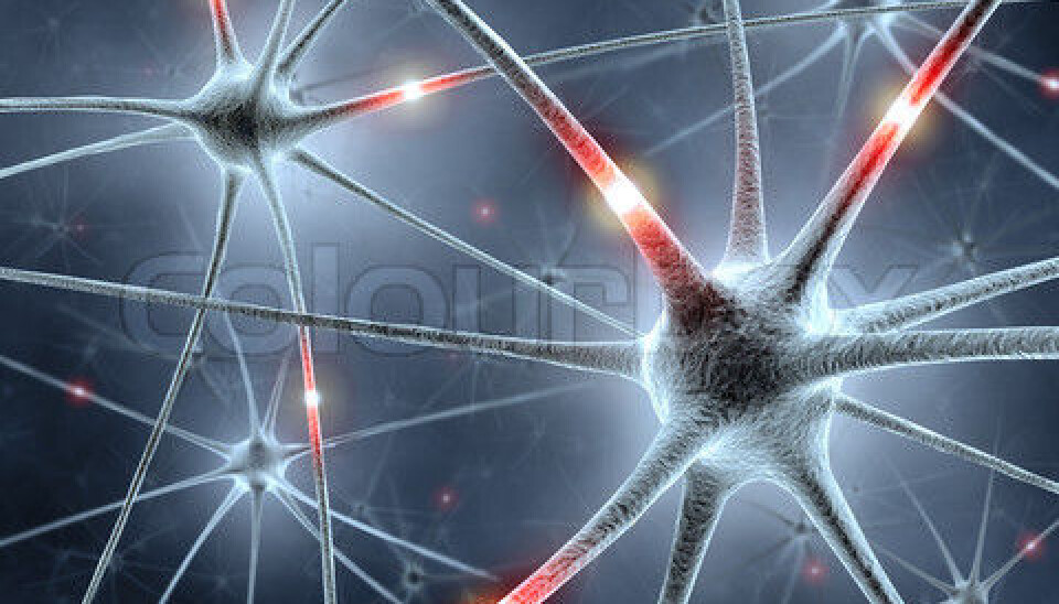
Synapses are points of contact where one nerve cell transfers signals to another. These convey everything you sense, experience, feel, think, say and remember.
But what is actually taking place in the synapse? This has long remained a mystery.
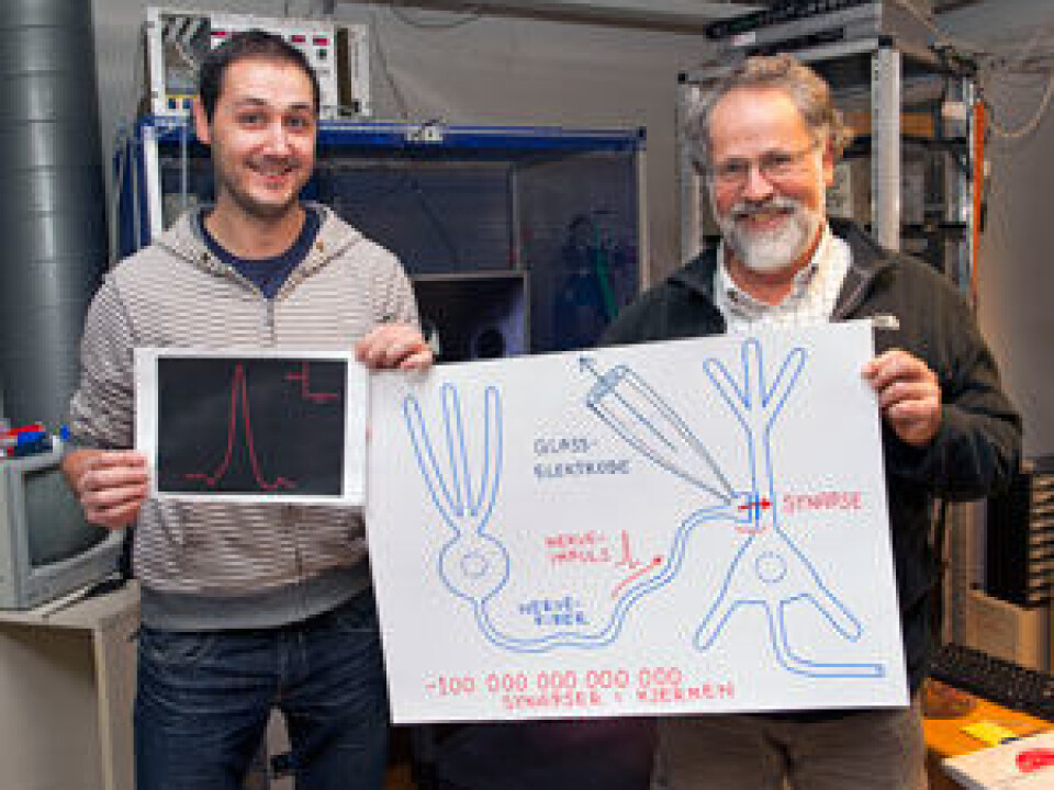
For the first time in Scandinavia, Pedro Mateos-Aparicio, researcher at the Institute of Basic Medical Sciences at the University of Oslo, has measured the nerve impulse in the end of a nerve fibre where it forms a synapse in the brain.
This will lead to greater knowledge about memory and learning, as well as about various disorders of the brain.
A complex system
Your brain contains more than 100,000 billion synapses. This is a staggering number, and says something about how microscopic these contact surfaces are – and about how complicated the brain is.
“Synapses have been studied in many different ways, but because synapses are so minute it is extremely difficult to measure the activity within them,” explains Professor Johan F. Storm.
Mateos-Aparicio has been working since last June to do just that. In November, he finally succeeded.
“By studying the cells’ behaviour at a more detailed level, we get a more precise view of the transfers,” he says.
A synapse is only a few thousandths of a millimetre in size. It goes without saying that measuring the activity that takes place within it is a challenging task.
The solution is a razor-thin glass tube.
A hole in the membrane
Using a micropipette, Mateos-Aparicio is able to create an opening in the membrane surrounding the synapse without it splitting.
Using a specially built machine, a thin glass tube is melted and stretched out to form a thin, hollow tip. The diameter of the tip of the micropipette is around one thousandth of a millimetre.
It is filled with a fluid resembling that found within the cell. The fluid in the pipette is able to conduct current and is connected to a transformer. The tip of the pipette is then guided down into the brain tissue towards the end of a nerve fibre where it forms a synapse.
To avoid any leakage, Mateos-Aparicio creates suction in the pipette so that the cell membrane attaches securely to its tip. Finally, he creates a hole through the membrane by generating a little extra suction.
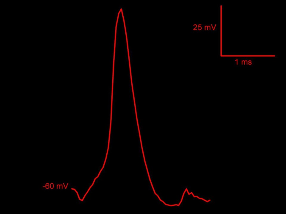
In this way, he is able to measure the nerve impulses within the end of the nerve fibre in the synapse.
The revealing signals
By measuring the activity in the end of the nerve fibre of the synapse, we can see the electrical signals that transmit information from cell to cell. This makes it easier to study in detail how synapses work.
It also allows researchers to find out more about what happens when we learn something new or memorise knowledge.
There is much evidence that memory is stored in synapses. When we use synapses and learn something new, the signal transfer in some synapses is a little more effective than previously. These changes can last for a long time.
Using the new method, the researchers can also see weaker electrical signals than those forming the actual nerve impulses.
“We suspect that the more subdued signals like these, which for many years have been overlooked by researchers, may also be important,” says Storm.
This is why we remember
There are many types of learning and memory.
The most important area of the brain for conscious memory is the hippocampus. This is where all the information that you remember is stored, and your life history is reflected in the changes in the synapses. When you watch a film, many synapses change. It is also due to the synapses that you can remember what you ate for breakfast this morning.
“The synapses in the hippocampus are probably absolutely vital for our conscious long-term memory – the things that we consciously know we remember and can talk about many minutes, hours, days and weeks later. The changes in the synapses of the hippocampus are probably the actual memory traces where this type of long-term memory is stored initially,” Storm explains.
Later, this type of memory is gradually transferred to the synapses in other parts of the cerebral cortex and becomes independent of the hippocampus. In other words, the synapses in other parts of the cerebral cortex are where our most long-lasting, conscious long-term memory is probably stored.
More knowledge of brain disorders
The hippocampus is part of the limbic system. This is a collective term for those parts of the brain that are old in terms of our evolutionary history. There is considerable activity here whenever you become angry, terrified or blissfully happy.
This system helps to regulate dopamine secretion, which is involved in controlling your emotional responses.
Given this, it is perhaps not so strange that researchers believe that changes to the synapses in the hippocampus are important causes of mental health disorders such as schizophrenia, as well as Alzheimer’s disease and epilepsy.
In this way, perhaps more detailed knowledge of synaptic activity may provide a greater understanding of these disorders.