THIS ARTICLE/PRESS RELEASE IS PAID FOR AND PRESENTED BY NTNU Norwegian University of Science and Technology - read more
The unsolved mystery of bad teeth in children
Many children have teeth that are practically falling apart due to weak enamel. Researchers have now studied whether a lack of vitamin D during pregnancy could be the culprit.
Tooth enamel is the body’s hardest substance, but some children have tooth enamel that is far from hard enough.
A study in the British Dental Journal reports that up to one in six children globally is affected.
Weak enamel is often discovered when baby teeth fall out, and the adult teeth start to erupt.

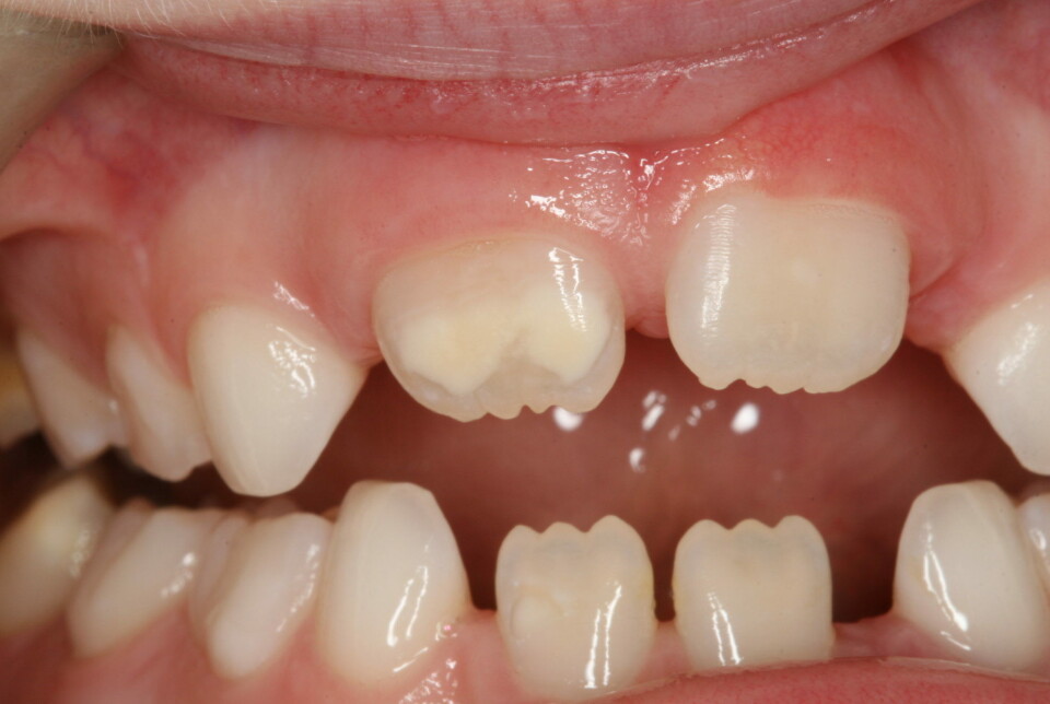
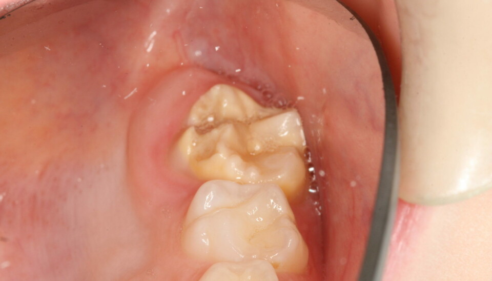
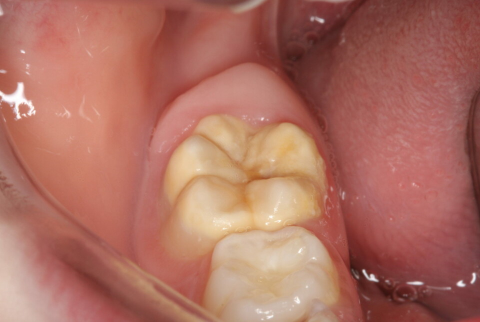
The weak enamel can appear as white and yellow-brown spots on molars and incisors in some children. In others, the teeth might be partly brown, with a rough surface. Some children are especially seriously affected and develop weak enamel on many teeth.
Even after many years of research, it is still not understood why some children develop enamel defects.
Sometimes the enamel is so weak that pieces of the teeth break off just by chewing normal food. In others, the enamel flakes off bit by bit.
Many children develop fear of dentists
Your child has a toothache. It hurts to eat, it hurts to brush the teeth, and it hurts to have them examined. All this pain can start a vicious circle.
Many children also develop a fear of the dentist. The pain of brushing teeth often leads to tooth decay. Caries occur because deposits accumulate that cannot be brushed away, exacerbating the problem.
Both parents, and sometimes the dentist, may think that the problems started with their child doing a sloppy brushing job – or maybe eating too much candy?
But that’s not the case.
The bad teeth are a result of the formation of enamel being disturbed.
The formation of enamel is a complicated process that takes place over many years, and begins during the foetal stage.
Strong enamel has lots of minerals in it, almost 99 per cent. Weak enamel contains few minerals, and more water and protein.
Have to pull adult molars at a young age
“It’s complicated to find treatment for these children. The fillings adhere less well and often fall out. Some children have to have their adult molars pulled at a young age because their teeth are in such bad shape,” Torunn Børsting says.
She is a PhD candidate at NTNU and project coordinator at the Dental Services Competence Center in Central Norway (TkMN).
As if all this is not enough, the teeth are also difficult to anaesthetise. An already painful tooth can thus become very painful to treat.
A researcher from Kings College in London has found that these children visit the dentist ten times more often than other children do.
Possible causes
So what can you do? Why is this happening? What preventive advice can be given?
Below are some of the possible connections that researchers in Norway and other parts of the world have ploughed through to try to understand why enamel development has been disrupted:
- Difficult birth, e.g. lack of oxygen or caesarean section.
- Infection in the early years of the child’s life.
- Use of antibiotics in the early years of the child’s life.
- Stress or illness in the mother during pregnancy.
- Lack of vitamin D in the mother during pregnancy.
- Low birth weight or premature birth.
- Environmental toxins disrupt development.
- Genetic factors.
Several studies have shown that illness in the first years of life can be linked to enamel defects.
However…
Many patients with enamel defects were not very ill, and others were often ill without developing enamel defects.
The figures for how many children are affected also fluctuate greatly, ranging from 2 to 40 per cent. Some people are of the opinion that the condition is more common now than in the past, but that is also unclear.
The enamel defect was first defined in 2001. Most of the studies have been carried since then.
In the past, many children had cavities in their first permanent (six-year) molars, and it might not have been that easy to determine that enamel defects were actually the problem.
“We simply don’t fully know the cause of this enamel failure. It’s incredibly frustrating because we can’t offer any preventive advice,” Børsting says.
Examined 176 pregnant women
Børsting recently published a study in which she checked vitamin D deficiency in the mother during pregnancy as a cause.
Vitamin D plays an important role in the absorption of calcium and phosphate in both teeth and bones.
The study was carried out over seven years. First, 176 pregnant women were examined twice during pregnancy to check their vitamin D levels. After seven years, the children were called in to check their teeth. They were between seven and nine years old when the dental health exams were carried out.
Børsting’s main theory was that if the children were born with a low level of vitamin D, it might be more difficult to increase the level enough after birth, as compared to children who start life with an adequate level of vitamin D.
The study showed that 32 per cent of the children had at least one six-year molar with enamel defects.
A connection was also found between the mothers who had low levels of vitamin D during pregnancy and enamel defects in children.
Still a mystery
Nine mothers withdrew from the study and did not have their vitamin D level measured during pregnancy – enough of them that a connection could not be statistically established.
“The frustration continues,” Børsting says.
Several studies have investigated the connection between vitamin D and enamel defects. In a study from Denmark, pregnant women were given supplements with vitamin D during pregnancy, which resulted in significantly fewer children developing enamel defects.
However, a study from the Netherlands, in which vitamin D was measured in the mother during pregnancy but which provided no supplements, did not show a connection between vitamin D and enamel defects.
The mystery continues. The conclusion will have to wait. Researchers just have to go back to the drawing board, apply for more funding and do more research.
References:
Almuallem, Z. & Busuttil-Naudi, A. Molar incisor hypomineralisation (MIH) – an overview, British Dental Journal, 2018. DOI: 10.1038/sj.bdj.2018.814
Børsting et al. Maternal vitamin D status in pregnancy and molar incisor hypomineralisation and hypomineralised second primary molars in the offspring at 7–9 years of age: a longitudinal study, European Archives of Paediatric Dentistry, vol. 23, 2022. DOI: 10.1007/s40368-022-00712-y
Silva et al. 'Etiology of molar incisor hypomineralization – A systematic review', Community Dentistry and Oral Epidemiology, 2016. DOI: 10.1111/cdoe.12229 Abstract.

Read more content from NTNU:
-
Researchers are surprised that these chemicals have skyrocketed in Svalbard
-
Why some people can’t stop pulling out their hair or picking their skin
-
Can Europe create AI that we actually understand?
-
Doctors and nurses believe their own substance use affects patients
-
When the offshore wind turbines fall silent: What happens to the waste?
-
“Norway used German soldiers to clear minefields and dump explosives in fjords and lakes"


