This article was produced and financed by The Research Council of Norway

When bone-eating cells gain the upper hand
Advanced osteoporosis is often the most severe sequela, or resulting condition, of plasma cell cancer. Abnormally functioning stem cells are a key causal factor.
Denne artikkelen er over ti år gammel og kan inneholde utdatert informasjon.
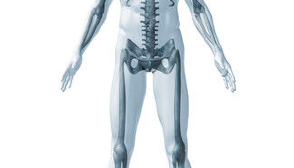
The human skeleton has the capacity to renew itself completely in the course of about ten years.
But maintaining strong, healthy bone structure requires the proper balance between cells that break down bone (osteoclasts) and cells that produce it (osteoblasts).
Cancer patients with painful bone conditions
“If the balance between osteoclasts and osteoblasts is disrupted, the bone may be weakened. This is precisely what occurs in patients with multiple myeloma, or plasma cell cancer,” explains Therese Standal, senior scientist at the K. G. Jebsen Center for Myeloma Research at the Norwegian University of Science and Technology (NTNU) in Trondheim.
For many cancer patients, the pain caused by destruction of bone is the worst consequence of their disease.
“These disorders are often painful and highly debilitating. Some patients may actually break a bone while lying down,” states Anders Sundan, head of the Jebsen Center and Professor in Molecular Cell Biology at the Department of Cancer Research and Molecular Medicine at NTNU, the same department where Standal is located.
Too few and immature cells lead to weak bone
Mesenchymal stem cells are stem cells found in bone marrow that have the capacity to develop into cells that produce everything from bone and fat to cartilage and muscle.
These stem cells play a key role in the development of the bone disorders suffered by patients.
Among other things, the researchers at NTNU have focused on understanding how bone cells develop from stem cells and how the skeleton breaks down in patients with multiple myeloma.
“It was previously believed that activation of osteoclasts, that is the bone-eating cells in the skeleton, was the main factor leading to an imbalance between bone degradation and bone construction. Our research shows that insufficient production of bone might be equally important,” explains Standal.
The researchers have discovered that many new bone cells stop developing before they reach full maturity, resulting in inadequate bone formation. These immature bone-forming cells have a high capacity to activate bone-degrading cells, creating a vicious cycle circle that results in a porous, fragile skeleton.
Molecules with a negative effect on bone mass
But why do the stem cells stop doing their job?
Standal and Sundan believe that the signal molecule hepatocyte growth factor (HGF) may hold part of the answer. Signal molecules take part in the transfer of information from a cell’s surroundings to the cell itself or between cells.
The HGF molecule is special in that it controls cell growth. It is active during normal cell growth but may also contribute to abnormal cell growth and the spread of cancer.
It has been known for 10-15 years that cancer cells can produce HGF. This is what occurs in malignant plasma cells, for example.
“Patients with bone marrow cancer and high concentrations of HGF in their blood have fewer mature cells producing bone than patients with lower concentrations of HGF do. In other words, a high concentration of this signal molecule has an adverse effect on the patient’s bone,” explains Standal.
Also the case for rheumatoid arthritis
Following up on this finding, Standal wanted to examine whether HGF also played a role in bone loss in connection with other diseases. In collaboration with researchers in Oslo and The Netherlands, she and her colleagues carried out a study on a large group of patients with rheumatoid arthritis.
“We found out that these patients also had high levels of HGF in their blood and poorer bone health than patients with lower concentrations of HGF,” she states.
Seeking new treatment methods
The Trondheim-based researchers believe that it will one day be possible to restrict HGF activity and thereby re-establish the balance between the cells that break down bone and those that build it.
“It will probably take some time before patients will be able to benefit from such treatment. At present, we are working on animal studies that we hope will lay the foundation for subsequent testing on human subjects,” concludes Sundan.
Translated by: Glenn Wells/Carol B. Eckman


