THIS CONTENT IS BROUGHT TO YOU BY the University of Stavanger - read more
Artificial intelligence could improve the quality of life for more patients following a stroke
Two researchers have teamed up to develop a tool that can help doctors make better and faster decisions when a cerebral stroke is suspected.
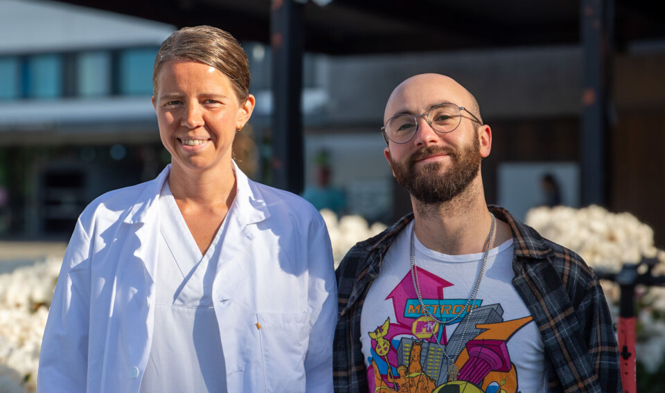
In her job as senior physician in radiology at Stavanger University Hospital (SUS), Liv Jorunn Høllesli has seen the importance of rapid and precise diagnosis of acute cerebral stroke.
Time is everything.
More precise imaging diagnostics
Images are taken of the brain almost immediately after the patient arrives at the hospital, and the images have a significant impact on the choice of treatment. But there are limitations to today's imaging diagnostics for acute strokes.
“A challenge in current imaging for cerebral stroke is limitations in distinguishing between brain tissue that can be saved and brain tissue that's already dead," says Høllesli.
Is it possible to develop a tool for even more precise image diagnostics than what is available today? Can artificial intelligence be used to improve the accuracy of the images?
Høllesli teamed up with computer scientist Luca Tomasetti. Together, they have come up with a solution that can provide faster and more precise diagnostics.
Differentiate between different types of tissue
The question the researchers asked was whether, with the help of machine learning, they could quickly find out exactly where the problem was. Machine learning is a method within artificial intelligence. And if the patient actually has a cerebral stroke?
Luca Tomasetti has been responsible for the technical solution for improved image diagnostics. His part of the twin project mainly involved developing new automatic methods for image diagnostics using machine learning.
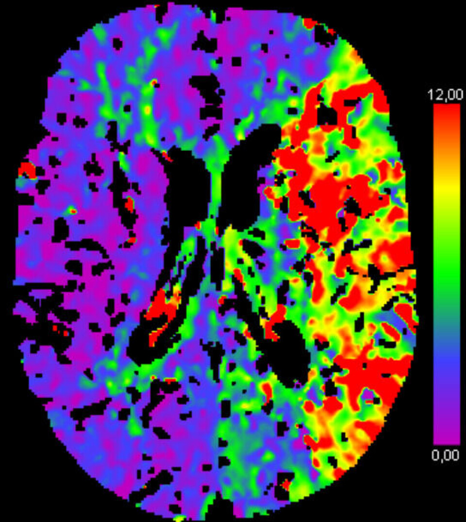
He has used images from CTP (computed tomography perfusion) scanning as input for an artificial intelligence network that can segment the areas of the brain with reduced blood supply.
In other words, identify the areas of the brain that should be treated.
“Every minute, millions of brain cells die during a cerebral stroke. Therefore, it's necessary to develop accurate, fast, and automatic solutions to identify the location and size of the area affected by cerebral strokes,” says Tomasetti.
Their research shows that using images based on CT perfusion as input for artificial intelligence increases the possibility of distinguishing the stroke-affected areas.
“The results can help radiologists make better and faster decisions for patients suspected of acute stroke,” says Tomasetti.
Quick decisions are needed in the healthcare system.
When a patient with a possible stroke enters the hospital, both early and correct treatment is crucial for the outcome - whether the patient survives or dies.
Two projects – same data
It has now been four years since the collaboration between Høllesli and Tomasetti began. They have both used the same data in their doctoral theses and have met weekly to discuss and analyse the results.
“Liv Jorunn is a radiologist, so she is an expert in the medical field. I helped her with the technical part of her project, and she helped me with the medical part of my project. We discussed articles, we discussed results, we discussed experiments. It was an active collaboration,” says Tomasetti.
He says that the first step was to collect as much data as possible. The data they used was images of the brains of possible stroke patients.
“We used the same images in the studies. Although we analysed them in different ways, we had opportunities to compare the results,” he says.
Høllesli says that it is not sufficient, when diagnosing a patient, to have good data and technical expertise to develop good algorithms.
“We also need medical expertise, to see that the answers are actually correct. That is what has been so exciting about the project," she says.
Høllesli explains that if they find a method that is technically perfect, it is not helpful if it is irrelevant to the patients.
"People with medical and technical expertise must work side by side to develop the best methods. It's absolutely necessary,” she says.
Is artificial intelligence the solution?
Is it easier for a machine to interpret images of brain tissue than for the human eye? Will the result be more accurate?
“We have not investigated the accuracy of the diagnosis when a radiologist looks at the images compared to the machine. But we find a good match between what the machine finds and what the radiologist finds,” says Høllesli.
There has been a major technological development in the healthcare system in recent years. However, new machine learning tools do not yet constitute a large part of everyday life in hospitals.
The tool the two have developed has not yet been put into use in Norwegian hospitals.
“It's a matter of money and human resources. But artificial intelligence is already part of the healthcare system. The question is how we should use the technology in a good way,” she says.
A small revolution
Høllesli mentions Bærum Hospital as an example (link in Norwegian). Last year, they adopted an artificial intelligence application for interpreting X-ray images.
When patients with suspected fractures come to the hospital, X-rays are taken. The images are processed and interpreted by artificial intelligence. The result is ready within a few minutes.
“It's a small revolution in diagnostics. Going from using a lot of energy and resources to interpret images, compared to using results from a machine,” she says.
Tomasetti also has high expectations for the use of artificial intelligence in healthcare.
“Of course, there are many challenges along the way, but there's also a lot of potential. As I see it, methods within artificial intelligence are perfectly suited to the healthcare sector. But we must allow the doctor's own interpretation and explanation of the results," says Tomasetti.
He further adds that doctors will still need to see the patient with their own eyes. New tools can probably make everyday work easier and contribute to improved diagnostics and treatment.
References:
Høllesli, L.J. Stroke Mimics and In Depth Analysis of Computed Tomography Perfusion in Patients with Acute Ischemic Stroke, PhD thesis at the University of Stavanger, 2024.
Tomasetti, L. 'Automatic AI-Driven segmentation of Acute Ischemic Stroke Regions with CT Perfusion Images', PhD thesis at the University of Stavanger, 2023. (Abstract)
———
Read the Norwegian version of this article on forskning.no

This content is paid for and presented by the University of Stavanger
This content is created by the University of Stavanger's communication staff, who use this platform to communicate science and share results from research with the public. The University of Stavanger is one of more than 80 owners of ScienceNorway.no. Read more here.
More content from the University of Stavanger:
-
“Our research can contribute to the development of batteries that store more energy"
-
How the Vikings protected themselves from attacks
-
Like tuning forks in space: A final pure tone reveals the mysterious interior of neutron stars
-
Could scented books encourage more kids to read?
-
Cathedral's lost treasures uncovered
-
Norwegian researchers can contribute to changing the Big Bang theory


