An article from Norwegian SciTech News at NTNU
A village of neuroscientists
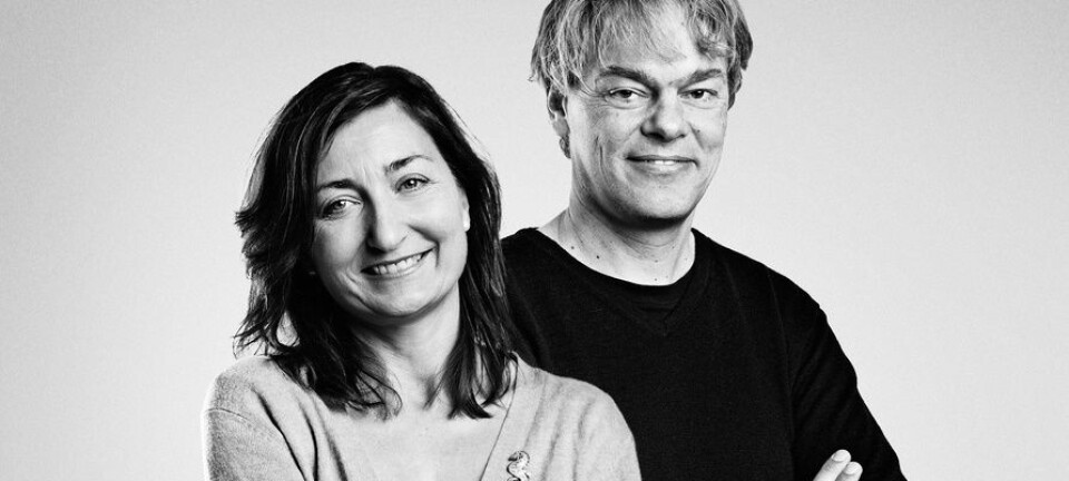
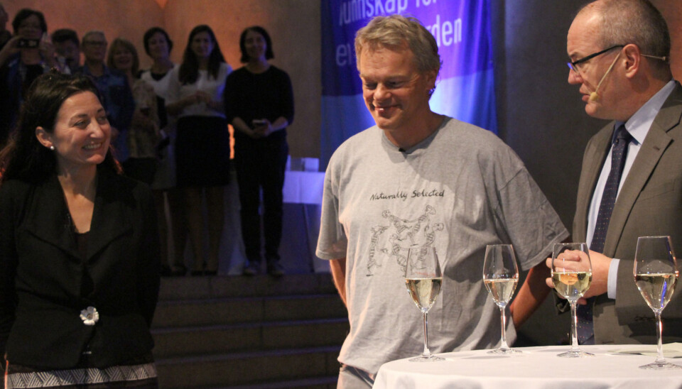
2014 NOBEL PRIZE: Nobel laureates May-Britt and Edvard Moser have spent the last two decades building a village of neuroscientists, molecular biologists, physicists and anatomists – whose different and complementary skills are key to the goal of unlocking the secrets of how the brain actually computes.
Denne artikkelen er over ti år gammel og kan inneholde utdatert informasjon.
The pioneering discovery of grid cells in the brain in 2005 by the Mosers and three of their graduate students shook the world of neuroscience to the very core – and is the reason the Mosers won the 2014 Nobel Prize in Physiology or Medicine with their former mentor, John O’Keefe. Their subsequent research showed how place and grid cells make it possible to determine position and to navigate.
Grid cells, the neurons that act like a GPS in the brain, gave neuroscientists an unexpected glimpse deep into the workings of the mind, where they could see how it made sense of the space around it.
“This is probably among the first instances where a process in the middle of the brain… has actually been understood in terms of what neurons are involved, what these neurons do, and to some extent how they collaborate,” Edvard Moser said in a recent interview. “The grid pattern is something that the brain has invented by itself, you don’t have any grid pattern in the outside world, so none of the senses conveys a grid to the brain. … In that sense it puts us on the track of finding out how the brain makes its own codes.”
In pursuit of this goal, and with the steady support of the Norwegian government, the university and the Kavli Foundation, the Mosers have built a laboratory of in-house expertise from disciplines as diverse as physics and molecular biology.
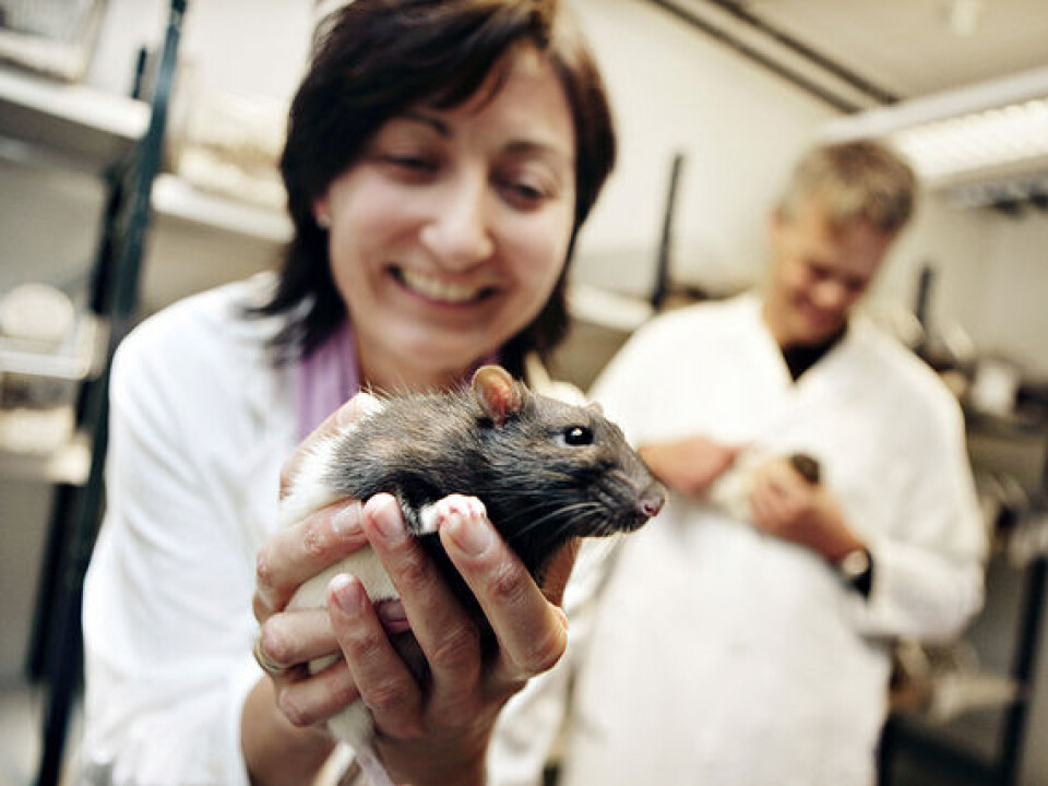
While researchers at the KI/CNC are divided into separate groups depending on their focus, together they are working towards a common goal – nothing less than cracking the brain’s codes, mapping its circuitry and figuring out how complex thoughts, such as memory, arise.
What follows is a peek at four of the research teams at the KI/CNC whose work complements the work of the Moser’s own (fifth and original) group. A sixth group from Belgium, headed by Emre Yaksi, will begin after the New Year.
A most complex organ
Group leader Cliff Kentros, who trained at NYU and Columbia and came most recently from the University of Oregon, reminds us that the brain is the most complex structure in the known universe, where every square millimetre of space is packed full of networks that enable us to think, feel and move. Kentros’s background is in molecular neurobiology.
“What is so different about the brain relative to all the other organs?” he says. “At the end of the day it is the anatomy, there is more anatomical complexity in even a tiny piece of the brain than there is in the rest of the body combined.”
Kentros says what neuroscientists need right now is the equivalent of a circuit diagram, so they can figure out how the brain actually works.
The brain “is an incredibly complex set of interconnections between these electrical connections we call neurons, another word for this is circuitry,” he said. “So if you are an engineer and want to figure out how a circuit works, the first thing you need is a circuit diagram.”
Here is Kentros’s contribution to the Moser village: he can create a variety of special transgenic mice that give neuroscientists the ability to perceive and control specific neural activity in selected parts of the brain – for example, in the medial entorhinal cortex, the part of the brain where the Mosers found grid cells.
In addition, he designs and uses viruses, such as a “defanged” rabies virus that acts like a tracer in the brain, because it has been engineered to jump from the first neuron it infects to just one and only one other neuron. The virus carries a protein that causes the neurons it infects to glow when researchers shine a special light on them. In this way, the researchers can see which neurons are talking to which other neurons.

“That’s what I bring,” he said. “The ability to make new kinds of mice and viruses… if you wanted to represent me as a mad scientist, it would be easy.”
These tools enabled Kentros and David Rowland, then a PhD and now a post doc in the Moser lab, to discover a shortcut in a key neural circuit in the hippocampus. They published their findings in 2013.
Now Kentros and colleagues are trying to understand the significance of this shortcut.
The transgenic mice also allow them to actually turn the activity of selected cells up and down, or on and off, to see what happens then.
These tools “allow us to do unprecedented things,” he said. “Most of the things we do are observations – you eavesdrop on the cells. But now we are into manipulation. What if cells fire more – does it matter? And it does it matter if they fire less? That tells us what kind of math the circuit does.”
Patterns and inference
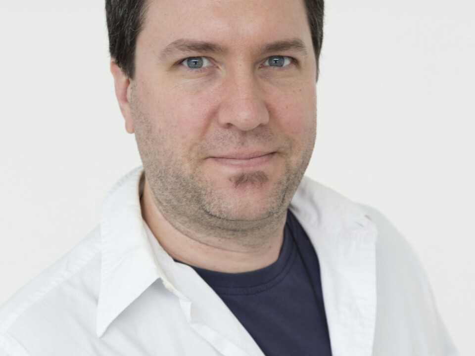
If Cliff Kentros is the neuroscience equivalent of an electrical engineer, mapping out the circuit diagrams of the brain, then physicist, professor and group leader Yasser Roudi is the “quant”, someone who specializes in handling huge amounts of data and extracting patterns and inferences from them.
“I am interested in theories of information processing,” Roudi said. “The brain is one representation of that, a powerful representation of an information processing machine that has been built through evolutionary processes.”
What interests Roudi most is “incomplete data where you want to infer something… in the most interesting cases, these data are stochastic (random), probabilistic, indirect observations of an underlying process, and you want to infer that underlying process.”
It’s not hard to see how this research passion can be applied to the information that neuroscientists in the Kavli labs collect when they attach measurement electrodes to their lab rats and let them freely roam in a box, searching for chocolate treats.
These experiments can record from as many as 186 electrodes simultaneously, but that is just a fraction of the number of neurons in a rat’s brain. Although it is possible to see patterns emerge from these data even with simple tools, the type of analysis offered by the work of Roudi and his group allows the researchers to dig much deeper in the data and to extract much more information from it.
A problematic interneuron
In addition to working on methods for statistical inference in complex data, Roudi also works on relating how the patterns that emerge from data are actually produced through the interactions between neurons in the nervous system, and specifically in the part of the nervous system that is involved in memory and navigation.
Roudi’s colleague Menno Witter likes to tell the story of how Roudi’s team was able to help explain some puzzling findings about the behaviour of stellate cells, star-shaped neurons that are found in the area of the brain where grid cells are found.

Witter’s then-postdoc, Jonathan Couey, wanted to understand more about how this area of the brain generates the characteristic firing patterns of grid cells, and spent two years recording information from the stellate cells in the area. But the data Couey collected didn’t seem to make any sense, Witter said.
“The bottom line was that stellate cells don’t talk to each other, they only talk to inhibitory interneurons, there is a neuron in between that inhibits them,” Witter explained. “So essentially if nothing happened in the network it would be dead silent, and that is weird.”
Witter has compared Couey’s initial puzzling findings to two people (the stellate cells) trying to have a conversation through a third person (the inhibitory interneuron). What Couey found would be the equivalent of every time one of the people tried to initiate a conversation, the person in between tried to stop it.
“It was crazy,” Witter said. “After two years of very hard work and recording from thousands of these stellate cells that was the conclusion, and we got stuck. So we decided to talk to Yasser, and said, ‘Can you check to see if it is feasible to have a network that would generate a grid cell firing pattern based on these principles?’”
It didn’t take long for Roudi’s group to see that the biological data could in fact generate a grid. But what came as even more of a surprise was when Roudi and his group realized that the same model could be used for explaining a puzzling set of data that had been collected several years earlier by one of the Moser group’s researchers. The result was that the researchers were able to publish two back-to-back articles in Nature Neuroscience in January 2013 explaining the connected findings.
Roudi says that kind of partnership – between a theoretical physicist like him, and experimental biologists, like the rest of the Kavli team – is a key aspect that makes the institute unique.
“Our interactions here are very bidirectional,” Roudi said. “And that is one of the unique things about having a theoretician here. The Mosers are very good experimentalists, but they are not only technically good, they listen. If I tell them I think you have to do this kind of measurement or study this, they are willing to invest their time and energy into it.”
The anatomist
Menno Witter was the first professor to join the Mosers at the then-Centre for the Biology of Memory in 2007, when he was appointed head of the functional neuroanatomy group. But the relationship between the Mosers and Witter extends to as early as 1990, when the Mosers were just beginning their academic career.
“My background is in comparative anatomy,” Witter said. “Give me a brain, any brain, and I will probably figure out what is where because I have done it all my life. I have looked at the brains of many different animal species, I have worked on guinea pigs, on rabbits, on cats, dogs, and tree shrews. I have worked on a lot of strange animals. Right now I am exploring the possibility of working with a consortium on bird brains.”
Witter’s deep knowledge of brain anatomy makes his expertise invaluable when researchers want to measure activity in specific parts of the brain but don’t know precisely where to find those parts.
For example, when researchers from the Weizmann Institute of Science in Israel wanted to measure activity in grid cells in bats, they contacted Witter for help finding just the right spot in the bat brain. One thing led to another, and “now I am working on an atlas to figure out the anatomy of the bat,” he said.
Witter’s lab looks for connections between different parts of the brain by using chemicals that are taken up by different parts of the neuron. This ability to visualize connections, combined with Witter’s knowledge of the brain, makes for a strong research relationship between Witter’s group and the Mosers’ group.
“I have my concept of how the brain is wired up,” Witter said. “And I try to convince the Mosers that (different projects) would be interesting to do. And they do projects where they ask me ‘how is this connected, how does this work, because we have these data and we don’t understand them’.”
Memory and decision making
For example, the trio has recently collaborated on research in an area of the brain that is important in connecting the decision making area of the brain to the memory system. The researchers chose this location because Witter knew lot about its connections.
When researchers disconnected the decision making area of the brain from the memory area in rats that had been trained to move in a certain pattern, the rats knew where they had come from, but couldn’t make the right decisions to move forward in the correct behavioural pattern.
The findings makes sense, Witter said, “because if we decide, we generally use our memory to realize what the consequences might be: I have been doing this in the past, so if I now decide to do this now, that is in line with my memory.”
“It is a beautiful result illustrating that by knowing a lot about the wiring you can make functional predictions,” he added.
For the future, Witter says he will work on explaining the differences between two parts of the entorhinal cortex.
These two areas, called the lateral and medial entorhinal cortex, “have the same name, they look similar, they have the same connections in the hippocampus, but there are a lot of subtle differences,” he said. It turns out these two areas are also very different with respect to their embryological cortical origin, he said.
“Now we have an interesting challenge,” he said. “How do two areas that are developmentally different end up so similar in terms of what they look like, while functionally, they are very different? So that is my challenge for the future.”
A hunt for mirror neurons
Jonathan Whitlock completed his doctorate at the Massachusetts Institute of Technology in 2006 and first came to Trondheim in 2007 as a post doc. He’s now the institute’s newest group leader – at least until Emre Yaksi comes to the institute in January – and will expand the scope of the Kavli Institute in two areas.
The first concerns a special type of motor neuron in the brain called a mirror neuron. First discovered in the early 1990s, these neurons fire both when an animal acts and when an animal sees another animal make the same action – which explains their name. They were discovered in macaque monkeys and were later found in humans and birds, and it is thought they are found in other social animals.
Whitlock was awarded a € 1.5 million start up grant in 2013 to look for these mirror neurons in rodents –and if he finds them, he can begin to map out the circuitry of how they relate to the rest of the brain.
“Bringing the study of mirror neurons to rodents would be a major step forward,” Whitlock said. “That means it would finally be possible to test whether mirror neurons are actually involved in the processes which they are believed to underlie – things like vicarious learning and social interactions, or whether they go awry in mental dysfunctions such as autism spectrum disorders.”
Planning and then moving
The second area where Whitlock will focus his efforts builds on his work as a post doc with an area of the brain called the parietal cortex. The Mosers, in contrast, have mainly focused on spatial mapping, with grid cells and other cells in the hippocampus and the entorhinal cortex.
The parietal cortex is an area of the brain that plays an important role in visual attention, working memory, spatial processing and movement planning. So Whitlock’s work involves navigation, but with twist.
“The thing you have to realize at the outset is that navigation is not something that just the grid cells or hippocampus does, the whole animal does it,” Whitlock said. “It is a composite contribution from the brain, it is complex behaviour. It’s not just using your eyes or your legs, the whole animal is navigating.”
To do this Whitlock will look at navigation from standpoint of serial motor planning, where the animal has to figure out how to get from point A to B. He’ll work with rodents and will record information from cells in the parietal cortex as the brain plans for movement.
It’s known that cells in the parietal cortex “have a sequential planning element to them – it precedes the movement by about a quarter of a second on average, but can extend more than one second ahead of the animal,” he said. “I’d like to see just how far into the future it is possible to predict an animal’s behaviour.”
One of the key things that Whitlock says he has learned from working with the Mosers is how to ask simple, direct questions “that let the brain tell you the answers,” he said.
“That is the fun thing about the brain,” he said. “There is so much stuff, if you just think about things in the right way, in a simple way. That is when you get at the neat stuff, if your question is simple enough. Otherwise it is easy to get caught up in your own contrivances and your own preconceptions.”
The letter on the wall
There’s a framed two-page letter – a fax, actually – next to Edvard Moser’s office door dated October 19, 1990 that begins “Dear Drs. Moser” – even though the couple were still master’s students at the time in the lab of Per Andersen at the University of Oslo.
The author was none other than Menno Witter, who wrote the fax as a young associate professor at Vrije University in the Netherlands. The Mosers had asked Witter if he thought was there a functional difference along the long length of the hippocampus, which forms a C-shaped structure in the brain. Their intention was to do a behavioural experiment on the structure by damaging one end or the other and then seeing what happened. But their first instinct was to reach out and ask questions before they began their work.
Witter had published a paper at the time that argued that there should be a difference along the structure, based on what he knew of the connectivity of the brain. He responded to the Mosers in his fax that he did not have much experience in behavioural studies, but described what he understood at the time of the brain’s networks in the area the Mosers wanted to study.
The Mosers went on to do the experiment, and “they actually found a difference,” Witter says. “I was very happy with that because they did the experiment I proposed that I couldn’t do because I didn’t have the facilities at the time.”
Witter says the letter is framed on the wall because the Mosers were amused by being addressed as PhDs, even though they were only master’s students.
“They were so proud because I addressed them as Drs Moser and Moser,” he said. “I didn’t know them and I figured they were PhDs because they worked with Per Andersen (at the University of Oslo) who to me was one of the godfathers of hippocampal work.”
Roudi also mentioned the fax. But like the theoretical physicist he is, he also saw a larger pattern in this one data point.
“It’s a beautiful letter,” he said. “You can see the mental process they were going through. It was theoretically framed, a step-by-step process. Biology is a descriptive field – it’s more common to say ‘we wonder what would happen if…’. But Edvard and May-Britt are the exact opposite. That’s the way they discovered grid cells. They have a 20-year history of asking the right questions.”


