THIS CONTENT IS BROUGHT TO YOU BY UiT The Arctic University of Norway - read more
This is how AI can contribute to faster treatment of lung cancer
It can lead to less costly examinations and more personalised cancer treatment.

Published
Lung cancer is one of the most widespread and deadly types of cancer in the world. In 2023, 3,319 Norwegians were diagnosed with lung cancer.
When the immune system detects cancer inside the lungs, it responds by sending out a group of immune cells. They attack the malignant cells in the tumour.
These immune cells are called tumour-infiltrating lymphocytes (TIL). They are an integral part of the body's battle against lung cancer.
Costly and time-consuming measurements
TIL cells can reveal how the cancer will develop. They can also indicate which treatment will work best.
Because of this, doctors examine tissue samples from the lungs under a microscrope. They map the TIL cells inside the tumour. The more cells they find, the better the prognosis.
The problem is that these examinations are very costly and time-consuming. The amount of TIL cells in the tissue samples may also be interpreted differently from doctor to doctor.

Researchers have now developed artificial intelligence (AI) that can make this task simpler. The technology can provide several benefits for the healthcare system – from less costly examinations to faster and more personalised cancer treatment.
“We sought to examine how machine learning can simplify this task. Now we know that it works pretty well,” says researcher Nikita Shvetsov at UiT The Arctic University of Norway. There, he participates in a machine learning group and the AI centre SFI Visual Intelligence.
Recognises healthy, diseased, and dead lung tissue
Machine learning is about enabling computers to learn something they did not know before. This is done by training them on large amounts of data. In Shvetsov's case, hundreds of digital images of lung tissue.
Parts of the dataset come from the University Hospital of Northern Norway. Researchers there are close collaborators in the researchproject.
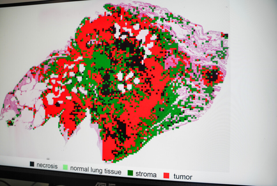
In this way, the AI system has learnt what lungs look like – including healthy, diseased, and dead lung tissue. This allows it to filter out image sections that don't contain tissue.
The technology falls under so-called computational pathology. It is a sub-field within digital pathology. Data-driven algorithms are used to analyse digital images of cells and tissue.
Shvetsov says this allows for more creative and automated ways of conducting these analyses.
“By using advanced algorithms and machine learning methods, the machines can find and draw out the most important sections from the images. This reduces the need for manual work and the potential risk of errors,” Shvetsov explains, adding:
“Not only does it contribute to more precise analyses, but it also enables doctors to make faster decisions that directly impact the patients.”
Done in a couple of minutes
But digital images are often large. So large that analysing the contents of a single whole slide image can take an entire day – even with AI.
One challenge is therefore to develop systems that use fewer resources in terms of computing power, electricity, and expensive, specialised graphics processors.
Shvetsov's AI system represents a different, yet more resource-efficient way of analysing such medical images.
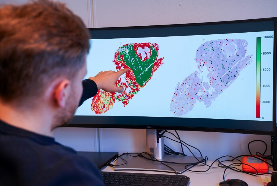
“A key feature of our system is that it doesn't analyse the entire whole slide image. In the first step, it selects small and random parts of the image to examine. Even though it only looks at a few sections, the system still provides a reliable overview of the number of TIL cells in the tumour,” he says.
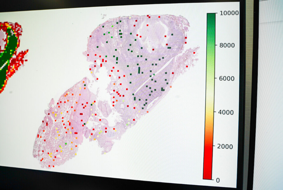
The next steps involve classifying image sections that contain TIL cells and mapping the cell quantity. This is done using an algorithm that recognises cell patterns in the image, counts different cell types, and identifies which of them are TIL cells.
The number is then converted into a score indicating the total concentration of TIL cells in different areas of the lung. Experiments from the study show that the scoring system provides an accurate indication of the patient's prognosis.
All of this is done in just a couple of minutes.
User-friendliness is important
Shvetsov emphasises that the score itself is not a sufficient basis for making an accurate prognosis.
The system must be able to show how it arrived at its conclusions. This transparency is essential for establishing trust in AI as a medical tool among doctors and patients.
“In other words, the technology must be able to provide doctors with visual evidence. Our system allows them to review which cells it has identified, segmented, and classified. This allows them to verify the results and confirm that the count is actually accurate,” he says.
To check this properly, such systems must be easy for doctors to use. User-friendliness is a central focus of the research project.
“Lack of technical expertise can make healthcare professionals reluctant to adopt and use AI systems. That's why it's important that both the system and its results make sense – even for a non-expert,” Shvetsov explains.
Adaptable to other cells and cancer types
Another goal of the project is to develop AI technology that can be adapted to other medical applications.
While the system was initially trained to map TIL cells, it can be adjusted to recognise other cell types that may also indicate the cancer prognosis.
The system can be fine-tuned to detect other biomarkers related to the body's immune response, such as PD-1 proteins.
"It can also be adapted for cancers other than lung cancer. Although this requires additional training data, integrating this into the system is quite straightforward,” he says.
Meant to assist doctors – not replace them
While the system is still far from being a finished product, Shvetsov's research lays the groundwork for a promising medical tool.
But does that mean doctors might one day be replaced by machines that do their job?
Quite the contrary, Shvetsov replies. He stresses that AI tools are meant to assist doctors in their clinical work. No matter how advanced AI becomes, it will always be necessary to have real human beings in the loop.
“As with other AI-based tools, the goal is not to replace healthcare professionals with automated machines. Our aim is to make their lives a little easier,” he says.
Reference:
Shvetsov et al. Fast TILs—A pipeline for efficient TILs estimation in non-small cell Lung cancer, Journal of Pathology Informatics, vol. 17, 2025. DOI: 10.1016/j.jpi.2025.100437
———
Read the Norwegian version of this article on forskning.no

This content is paid for and presented by UiT The Arctic University of Norway
This content is created by UiT's communication staff, who use this platform to communicate science and share results from research with the public. UiT The Arctic University of Norway is one of more than 80 owners of ScienceNorway.no. Read more here.
More content from UiT:
-
Stomach ulcers: Better treatment on the horizon
-
What affects a chatbot's ability to solve logical problems?
-
AI can help detect heart diseases more quickly
-
Why does Norway need its own AI law?
-
Researchers reveal a fascinating catch from the depths of the sea
-
How can we protect newborn babies from dangerous germs?


